Exposure To Food Increases Brain Metabolism
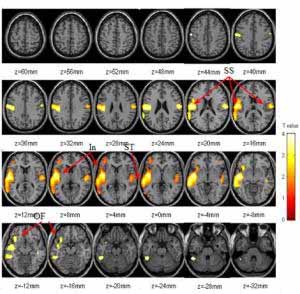
This figure shows the areas of the brain that "lit up" with higher metabolism during food stimulation in the study. The colored areas are from a statistical parametric mapping (SPM) analysis of PET scans of all subjects in both the food stimulation and neutral intervention conditions of the study. These images are superimposed on MR images from one person's brain (taken from above), which are used to demonstrate correct anatomical positioning. All "lit" areas represent significant differences in metabolism between the two study conditions: white represents higher metabolism while red represents lower metabolism. Most intriguing is that the orbitofrontal cortex (see arrows in the two lower left brain images labeled "OF") has higher metabolism. Studies at Brookhaven and elsewhere have shown that this brain region, which houses the dopamine system, is also active in addiction, reward and motivation.
(Image courtesy Brookhaven National Laboratory)UPTON, NY -- Scientists at the U.S. Department of Energy’s Brookhaven National Laboratory have produced new evidence that brain circuits involved in drug addiction are also activated by the desire for food. The mere display of food — smelling and tasting favorite foods without actually eating them — causes increases in metabolism throughout the brain. Increases of metabolism in the right orbitofrontal cortex, a brain region that controls drive and pleasure, also correlate strongly with self-reports of desire for food and hunger.
Brookhaven scientists have conducted previous research showing that the right orbito-frontal cortex is involved in compulsive behaviors characteristic of addictive states, and that this brain region is activated when addicted individuals crave drugs such as cocaine (see related story). They have also shown that food stimulation, as done in this study, increases levels of dopamine, a neurotransmitter involved in pleasure and reward, in the brain’s dorsal striatum (see related story). Additionally, to better understand the relationship of the dopamine system to obesity, they looked at the brain circuits of obese individuals and found that, like drug addicts, these individuals had fewer dopamine receptors than normal control subjects (see related story).
In the new study, the scientists looked at how 12 food-deprived, normal-weight study volunteers responded to their favorite foods by using positron emission tomography (PET), a brain-scanning technique, to measure brain metabolism during food stimulation. Each volunteer was given an injection containing a radiotracer, a radioactive chemical “tag” designed to “light up” activated areas of the brain. The PET camera picks up the radioactive signal and measures the level and location of the tracer. Study subjects’ brains were scanned twice over a two-day period, with and without food stimulation. Subjects were asked not to eat for 17 to 19 hours prior to the study.
Volunteers were presented with foods that they had reported as their favorites on a pre-study questionnaire. The food was warmed to enhance its smell and the subjects were allowed to view and smell it, as well as taste a small portion placed on their tongues with a cotton swab. They were also asked to describe the foods as they tasted them. As a control, during scans when food stimulation was not used, subjects were asked to describe in great detail their family genealogy while they were presented with non-food related items. They were allowed to smell these items and they had a cotton swab dipped in water placed on their tongues to mimic the food-stimulation condition. Study participants were also instructed to describe, on a scale of 1 to 10, whether they felt hungry or desired food at the start of the study and at five-minute intervals for a total of 45 minutes.
The researchers found that food stimulation significantly increased whole brain metabolism. Metabolism was higher in all regions of the brain examined, except for the occipital cortex, which controls vision and would not be affected. The areas most affected were the superior temporal, anterior insula, and orbitofrontal cortices. Food stimulation also resulted in increases in self-reports of hunger and desire for food. Increases in metabolism in the right orbitofrontal cortex were the ones that were most significantly correlated with increased reports of hunger.
Betel nut
Low-fat blues
cacao-chocolate.com
cognitive-enhancers.com
Vitamins and mood
Pharmacogenomics
Docosahexaenoic acid
Catecholamine depletion
Mood, food and cognition
Nutmeg seeds and anxiety
Bad moods and sick hearts
PUFA deficiency and allergies
Refs
HOME
HedWeb
BLTC Research
The Good Drug Guide
Paradise-Engineering
The Hedonistic Imperative
MDMA: Utopian Pharmacology